
Radiographic assessment of the geometry and orientation of the hip joint involves a variety of measurements. In particular, for the assessment of possible dysplasia, that is, of the degree of coverage of the femoral head by the acetabulum, one of the measurements used is the centre – edge angle, or the angle of Wiberg (Wiberg’s center – edge angle). Assessment of this angle is a very good indicator of the shape of the acetabulum and is widely used in the treatment algorithms for disorders of the hip.
The Wiberg centre – edge angle represents the superolateral coverage of the femoral head in the coronal plane, as it appears on the anteroposterior radiograph of the hips. It is used to quantify developmental dysplasia of the hip, severe forms of which may lead to degeneration and destruction of the joint, that is, osteoarthritis, at a younger or older age, depending on severity. Small values of the angle indicate more severe dysplasia, while larger ones indicate increased coverage of the femoral head.
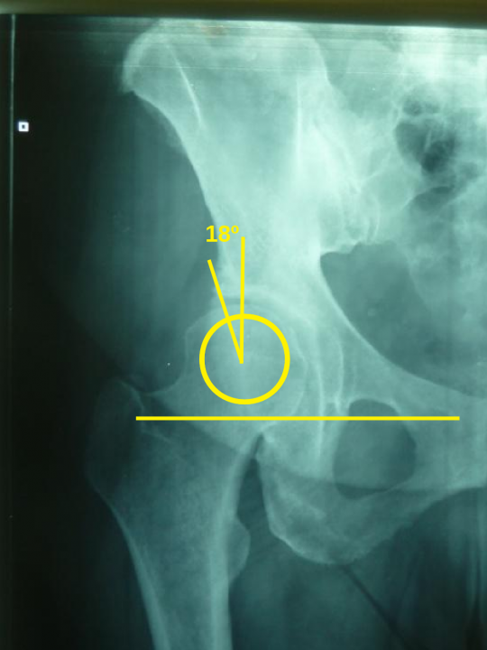
Wiberg described the angle that bears his name in 1939. According to his publication, this angle is formed by the line that begins at the centre of the femoral head and runs parallel to the axis of the patient’s body, and a line running from the centre of the femoral head to the lateral edge of the roof of the acetabulum (Fig. 1). According to Wiberg, angles greater than 25º were rarely associated with early osteoarthritis of the hip, those smaller than 20º (Fig. 2) were usually associated with early osteoarthritis, while angles between 20º and 25º were described as borderline.
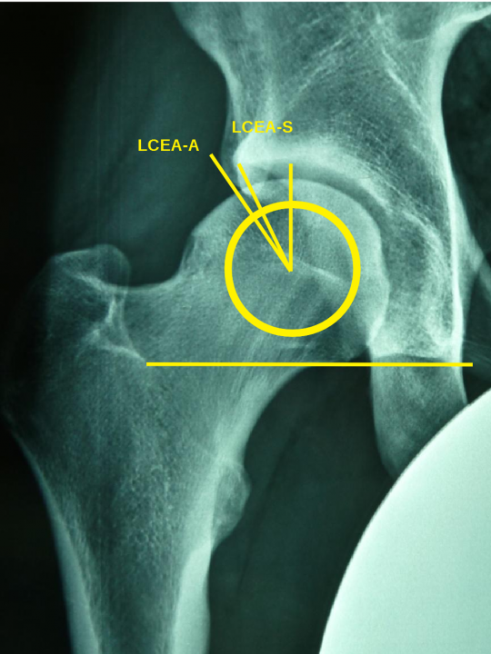
Several decades later, Ogata et al argued that, in paediatric patients in particular, this angle should not be measured to the lateral edge of the acetabulum as a whole, but to the lateral edge of the acetabular eyebrow (sourcil). The reasoning was that the radiographic lateral edge of the acetabulum, beyond the sourcil, does not contribute to load bearing, and so should not be counted in the total weight-bearing surface, and including it may lead to underestimation of the dysplasia. This observation appears to apply in young children, in posterior tilt of the acetabulum (retroversion) and in a low position of the anterior inferior iliac spine (AIIS). The angle was therefore subdivided into two separate angles, LCEA-S (Lateral center edge angle – sourcil) and LCEA-E (lateral center edge angle – edge of acetabulum), with the current literature supporting the greater reliability of the former (Fig. 3).
The clinical use of this angle has developed further still, since values greater than 40º are regarded as characteristic of overcoverage of the femoral head by the acetabulum, while the anterior centre edge angle (anterior center edge angle) is now also used. It is measured on the lateral radiograph (false-profile) and demonstrates anterior loss of coverage by the acetabulum at values below 20º, or overcoverage at values above 40º, which are found in cases with posterior tilt of the acetabulum and global or focal overcoverage of the femoral head by the acetabulum (global or focal anterior acetabular overcoverage).
References
- Wiberg G (1939) Studies on dysplastic acetabula and congenital subluxation of the hip joint: with special reference to the complication of osteoarthritis. Acta Chir Scand 83:53–68
- Ogata S, Moriya H, Tsuchiya K et al (1990) Acetabular cover in congenital dislocation of the hip. J Bone Joint Surg Br 72:190–196
- Petersen BD, Wolf B, Lambert JR, Clayton CW, Glueck DH, Jesse MK, Mei-Dan O. Lateral acetabular labral length is inversely related to acetabular coverage as measured by lateral center edge angle of Wiberg. J Hip Preserv Surg. 2016 Feb 29;3(3):190-6
- Lucas A. Anderson, MD; Jeremy Gililland, MD; Christopher Pelt, MD; Samuel Linford, MD; Gregory J. Stoddard, MPH; Christopher L. Peters, MD Center Edge Angle Measurement for Hip Preservation Surgery: Technique and Caveats Orthopedics. 2011;34(2):86

