

Injury to the acetabular labrum is a very common cause of chronic, persistent pain in the hip in young as well as older patients. It is seen mainly in people with intense sporting activity involving a large range of movement of the hip joint, such as martial arts, dance and ballet, but also football and basketball. It is very often combined with the condition of femoroacetabular impingement in which the abnormal shape of the femoral head or of the acetabulum creates an abnormal pattern of hip movement and an accompanying overuse syndrome.
In the early stages of the condition treatment is conservative, with analgesics, anti-inflammatory drugs and physiotherapy. When the symptoms are not brought under control, however, the most capable treatment for the problem is hip arthroscopy. Arthroscopic treatment of a labral tear not only relieves the symptoms but also has a protective role in preventing further wear of the joint, that is, osteoarthritis.
The labrum of the acetabulum has attracted a great deal of interest from hip surgeons in recent years, since it has been shown to be closely bound up with the overall health of the joint and with the onset of the first lesions of osteoarthritis.1 Its important role in the biomechanics of the joint is now generally accepted, and for that reason great emphasis has been placed in the international literature on the concept of surgical preservation of the labrum (preserving surgery) which usually means arthroscopic selective partial excision (partial labrectomy) or its repair and stabilisation (labral repair). 2
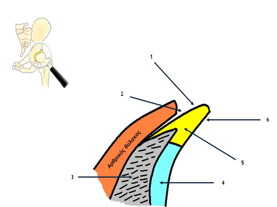
Figure 1: Vertical cross-section of the acetabular labrum, magnified
(1: capsular surface of the labrum, 2: perilabral sulcus,
3: bone of the acetabulum, 4: articular cartilage,
5: body of the labrum, 6: articular surface of the labrum).
Structure and biomechanical characteristics
If the acetabular labrum is examined under magnification, it will be seen that it is essentially the anatomical continuation of the articular cartilage (Figure 1). The articular cartilage – labrum complex therefore acts as a single surface for correct congruity between the femoral head and the acetabulum. For this reason, any damage to the labrum also has an effect on the structure of the articular cartilage and, secondarily, on the overall function of the joint.
In vertical cross-section the labrum appears triangular in shape, with its base attached to the bony rim of the acetabulum and to the articular cartilage, while its apex lies free. In many cases part of the bony attachment extends deep into the body of the labrum. It has two surfaces, the articular, which is in continuous contact with the femoral head, and the capsular, which forms the perilabral sulcus together with one side of the capsule.3
The labrum is fibrocartilaginous in composition. Studies have shown that it consists of dense bundles of collagen fibres, resembling a ligament histologically. Chondrocytes have been observed on the articular surface, and fibrocytes within its body.4
The boundary between the articular surface of the labrum and the articular cartilage, the so-called transitional zone, is the most vulnerable point of the inner surface of the acetabulum, and for that reason it sustains injury perhaps more than any other area.5
Its arthroscopic appearance varies from person to person, both in dimensions and in shape. The anterior labrum, however, always shows fundamental morphological differences from the posterior one, being broader and thicker, and it is also more vulnerable to tears (Figures 2 and 3).
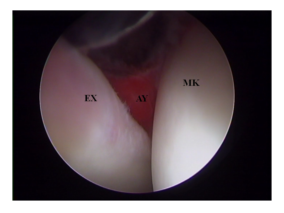
Figure 2: The anterior labrum is usually
quite thick and of greater width.
In most cases it cannot be distinguished
macroscopically from the articular cartilage
(ΕΧ: labrum, ΑΥ: synovial membrane, ΜΚ: femoral head).
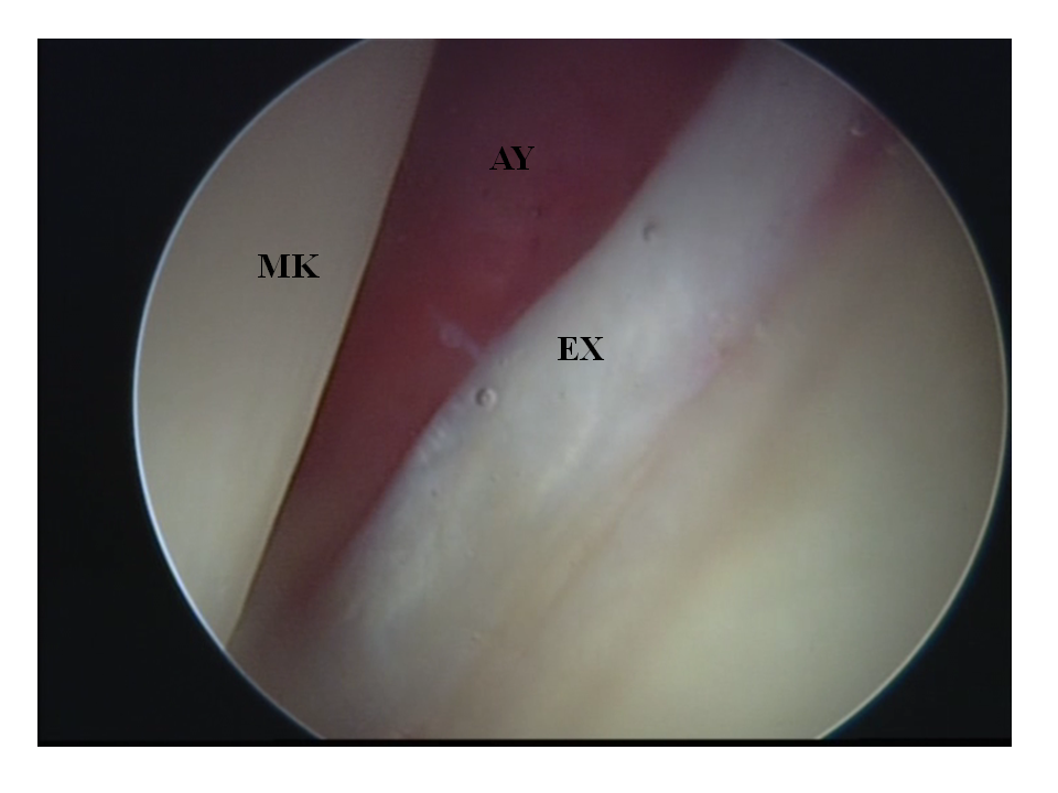
Figure 3: The posterior labrum is usually slender,
of small thickness, and is clearly distinguished from the articular cartilage of the acetabulum
(ΜΚ: femoral head, ΑΥ: synovial membrane, ΕΧ: labrum).
The biomechanical importance of the acetabular labrum has been described extensively in the literature.6 It appears to contribute to the stability of the joint by providing a seal (sealing effect), creating negative pressure within the joint, maintaining a continuous layer of synovial fluid and distributing loads over a larger surface.
Tears of the acetabular labrum
The recognised causes of a labral tear are femoroacetabular impingement of the cam or pincer type, the microinstability of the hip, an injury (an isolated traumatic episode with no underlying abnormal anatomy) and generalised degeneration of the joint. 7
The topography of the micro-anatomy in tears of the acetabular labrum is of particular interest. According to one classification, tears are divided into Type I, in which the articular cartilage separates from the labrum, and Type II, which are found at the free edge of the labrum (Figures 4A, 4B, 5A, 5B, 5C and 5D).8 Tears of the first category have been shown to be the more serious, that is, those occurring in the transitional zone between labrum and articular cartilage, since they extend deep as far as the subchondral bone, while secondarily they also produce detachment of the adjacent cartilage flap of the acetabulum.
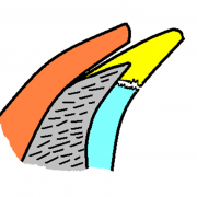
Figure 4A: Type I:
the tear has occurred in the transitional zone
between labrum and articular cartilage of the acetabulum.
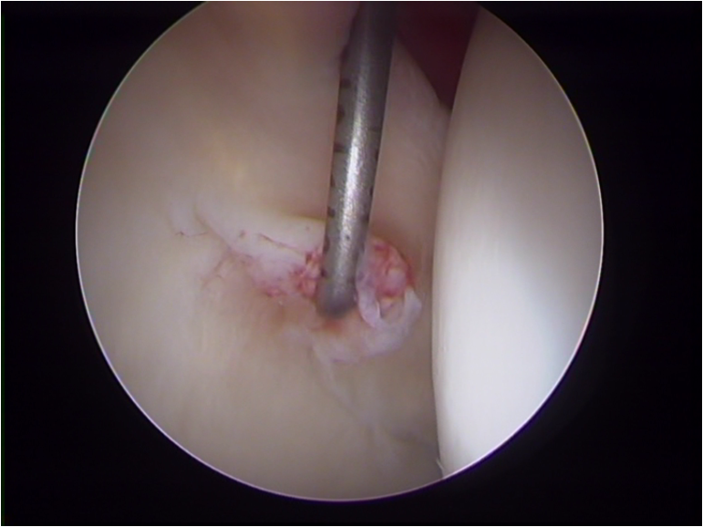
Figure 4B: The type I tear as seen arthroscopically.
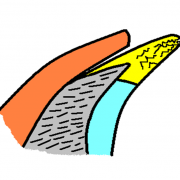
Figure 5A: The type II tear
involves multiple planes
within the body of the free edge
of the acetabular labrum
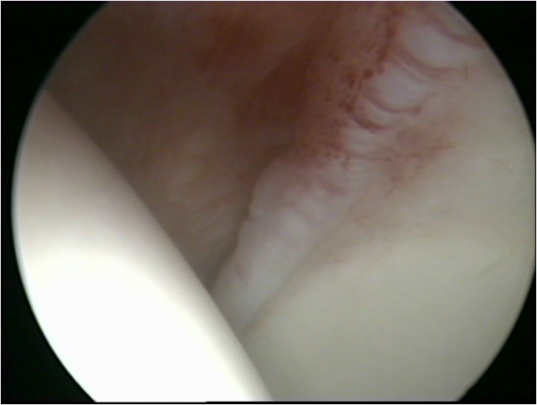
Figure 5B: An early-stage type II tear (fibrillation).
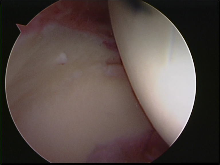
Figure 5C: A type II tear
involving the whole body of the labrum.
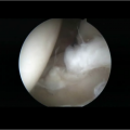
Figure 5D: An advanced type II tear
with complete disorganisation of the structure of the labrum.
Type I tears are the ones that, over time, produce progressive damage to the hip joint, since the detachments usually advance over a greater area, stripping bare wider regions of the subchondral bone (Figures 6A, 6B, 7A and 7B).
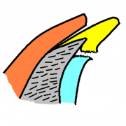
Figure 6A: The type I tear
has widened and detachment
of the labrum and of the articular cartilage
from the subchondral bone has begun.
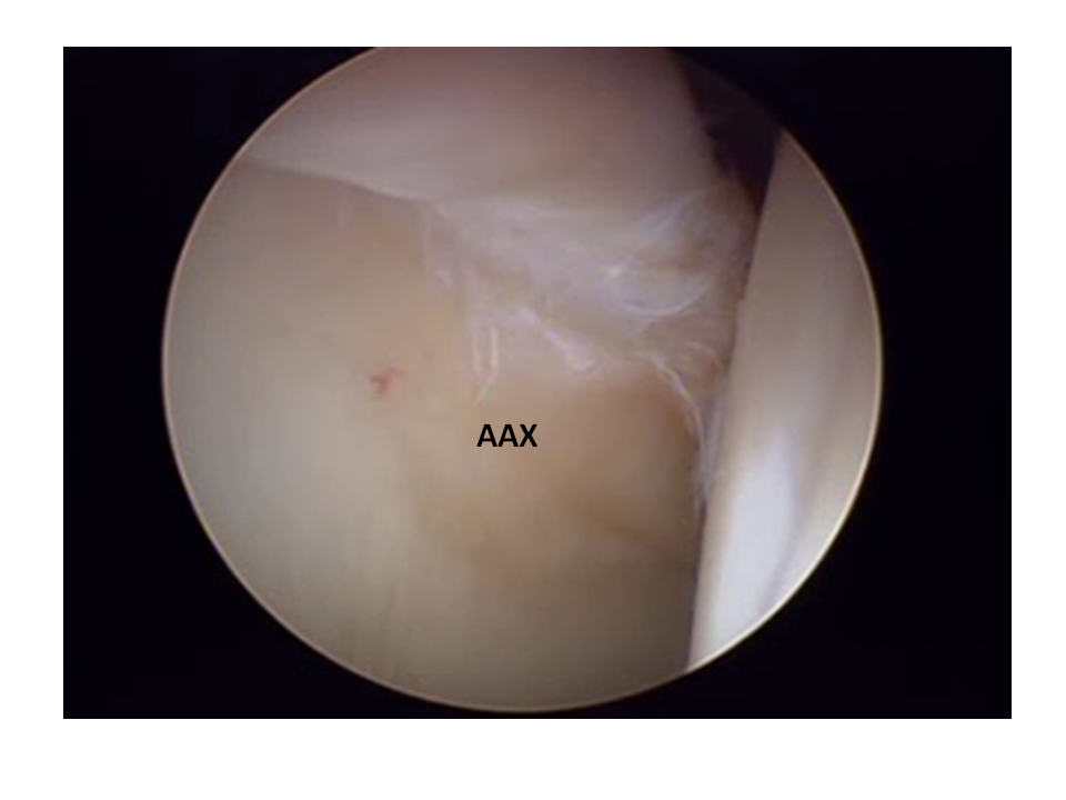
Figure 6B: A type I tear -
the detached articular cartilage (ΑΑΧ),
which has not, however, displaced,
appears in a darker colour.
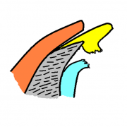
Figure 7A: A neglected labral tear -
both the labrum and the articular cartilage
have detached over a large area,
leaving an exposed surface of subchondral bone.
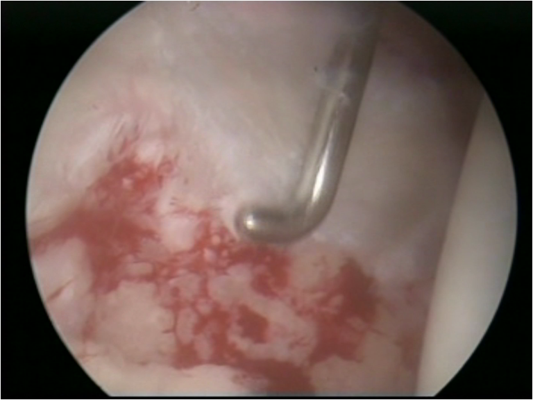
Figure 7B: Extensive detachment of the articular cartilage -
a grade IV chondral lesion on the Outerbridge classification.
The labrum is preserved,
but is completely detached at its base.
These extensive detachments are in effect the first lesions of degeneration of the joint, that is, of osteoarthritis of the hip, which may not be visible on radiographs or even on MRI, other than as mild, early osteoarthritis-type lesions (the appearance of small subchondral cysts, focal thinning of the articular cartilage).
Treatment of a labral tear
Conservative treatment with anti-inflammatory drugs, rest and physiotherapy always has a place, especially in the first stages in which symptoms appear. 9 It has been shown, however, particularly in the athletic population, that a return to the level of sporting activity that preceded the tear brings the symptoms back. In these cases, where conservative treatment has failed, surgical treatment is recommended.
The current method of choice for surgical treatment is the arthroscopic technique by which selective partial excision of the labrum, repair and stabilisation, or reconstruction is carried out. 10 The traditional technique of selective partial excision has shown good results over a decade. 11 With the ascendancy of the rationale of hip-salvage and hip-preserving surgery, however, the international trend is now to try to preserve the torn labrum, where that is feasible, by repairing and stabilising it with suture anchors (Figures 8A and 8B). The aim is to preserve this valuable anatomical structure, the labrum, with the ultimate goal of slowing the wear of the joint.
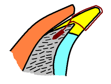
Figure 8A: The suture anchor has been placed
in the bony rim of the acetabulum,
and through the loop of the suture
the labrum is secured.
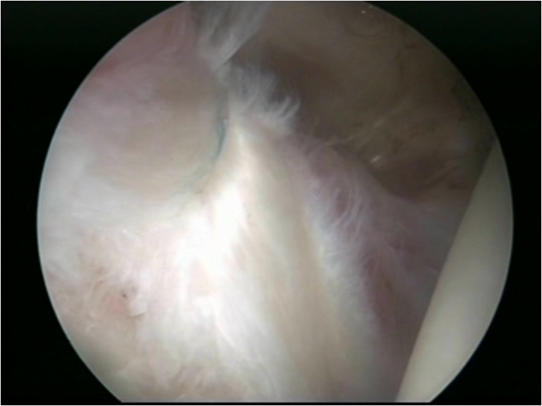
Figure 8B: Fixation of the labrum
with a suture anchor and sutures.
In cases with large detachments and chondral defects the use of the microfracture technique is recommended, either to attempt to stabilise the existing cartilage flap or for the potential production of fibrocartilage from the subchondral bone (Figures 9A and 9B).
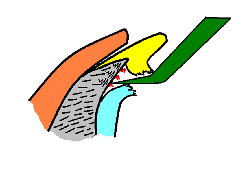
Figure 9A: With a special instrument (microfracture awl)
the microfracture technique is performed
in order to release pluripotent blood cells
that will help the cartilage flap to reattach.
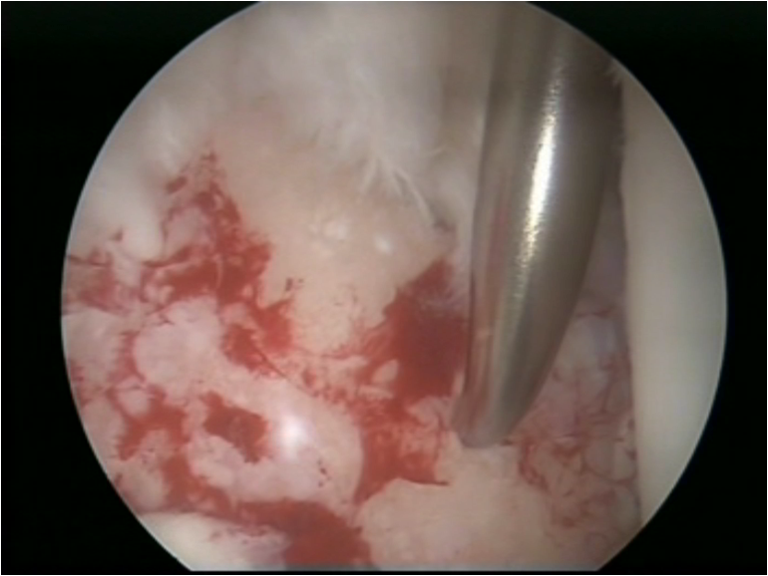
Figure 9B: In large chondral defects
the microfracture technique is used
to produce fibrocartilage.
In recent years reconstruction has emerged as an alternative surgical treatment in patients in whom the labrum is absent or extensively destroyed, and when it is not amenable to repair. Grafts of iliotibial band, gracilis tendon and ligamentum teres have been used in reconstruction. The first results of the technique appear encouraging. 12
Conclusions
The acetabular labrum has come to be recognised as a structure of great importance to the hip joint, both for its proper function and for preventing its early wear. Tears of the labrum may be the first stage in further structural damage to the joint. The most recent data in the current literature suggest that the view that every effort should be made to preserve it, either by the repair technique or by reconstruction, is gaining ground.
References
-
McCarthy JC, Noble PC, Schuck MR, Wright J, Lee J. The Otto E. Aufranc Award: The role of labral lesions to development of early degenerative hip disease. Clin Orthop Relat Res. 2001 Dec;(393):25-37.Bsat S, Frei H, Beaulé PE. The acetabular labrum: a review of its function. Bone Joint J. 2016 Jun;98-B(6):730-5. - Seldes RM, Tan V, Hunt J, Katz M, Winiarsky R, Fitzgerald RH Jr. Anatomy,histologic features, and vascularity of the adult acetabular labrum. Clin Orthop Relat Res. 2001 Jan;(382):232-40.
- Walker JM. Histological study of the fetal development of the human acetabulumand labrum: significance in congenital hip disease. Yale J Biol Med. 1981 Jul-Aug;54(4):255-63.
- cCarthy JC, Noble PC, Schuck MR, Wright J, Lee J. The watershed labral lesion: its relationship to early arthritis of the hip. J Arthroplasty. 2001 Dec;16(8 Suppl 1):81-7.
- Tzaveas Α, Villar R: Acetabular Labral and Chondral Pathology. The Open Sports Medicine Journal. 2010 Volume 4, pp 64-74.
-
Lage LA, Patel JV, Villar RN. The acetabular labral tear: an arthroscopic classification. Arthroscopy. 1996 Jun;12(3):269-72. -
Seldes RM, Tan V, Hunt J, Katz M, Winiarsky R, Fitzgerald RH Jr. Anatomy, histologic features, and vascularity of the adult acetabular labrum. Clin Orthop Relat Res. 2001 Jan;(382):232-40. - Bsat S, Frei H, Beaulé PE. The acetabular labrum: a review of its function. Bone Joint J. 2016 Jun;98-B(6):730-5. doi: 10.1302/0301-620X.98B6.37099. Review.
- Harris JD. Hip labral repair: options and outcomes. Curr Rev Musculoskelet Med. 2016 Dec;9(4):361-367. Review.
- Byrd JW, Jones KS. Hip arthroscopy for labral pathology: prospective analysis with 10-year follow-up. Arthroscopy. 2009 Apr;25(4):365-8.
- Ayeni OR, Alradwan H, de Sa D, Philippon MJ. The hip labrum reconstruction: indications and outcomes--a systematic review. Knee Surg Sports Traumatol Arthrosc. 2014 Apr;22(4):737-43.


